
Microscopic Mechanised Endodontic Root Treatments


Endodontic root treatment Berlin
Root canal treatment, endotontic treatment, endodonty generally only means that the rather wide diagnosis of “living tooth” or “dead tooth” can be treated mainly under differential diagnosis aspects. Initially, clinical findings and X-ray presentations are correlated to the anamnesis. The presentation of a tooth in the digital low-radiation X-ray or DVT shows further findings. First, the digital image in an editable file shows whether the bony and connective-tissue environment of the tooth shows any changes to naturally healthy or treated and healthy situations. These may be findings we interpret as ancylosis, inflammation, fistula, cysts, calcifications and many others. These findings of the dental environment form the basis of examination of the tooth and its inner canal system from the outside in. Now the tooth surface is assessed. This should also be similar or comparable to the neutrally healthy or treated healthy situation. Inner or outer inflammation consequences or changes to the inner or outer milieu may lead to contour changes that offer further diagnosis and prognosis indicators. Clinical microscopic differentiation of the findings on the tooth in question can lead to the following general classification, which then leads to a decision for further therapy measures. Smooth transfers are possible, because every finding and its description are only a momentary still image. Also see: BERLIN-KLINIK Index.
Highest technical service
It is important to us at BERLIN-KLINIK Zahnklinik Mitte to offer service at the highest technical and diagnosis levels in the disciplines offered. BERLIN-KLINIK ENDODONTIE even now meets standards that other places still philosophise about. Why? Among others, because our patients do not sporadically request higher-quality services but continually. We are equipped and ready for that.
Root Treatment Information:
Teeth that are untreated, clearly vital, pain-free, not loosened, not sensitive to tapping and completely without findings may show unexpected opening of the nervous hollow pulp with discharge of bright red arterial blood during therapy. If proper coverage of the wound under coffer dam is insufficient, proper root treatment promises success here. At BERLIN-KLINIK Zahnklinik Mitte, magnifying glass, surgical microscope or dental microscope are used first to prepare the canal system under view using flexible titanium files and special endodontic ultrasound files or diode lasers. The result is widely disinfected and then permanently closed. Root Treatment Surgical microscope. teeth that are vital, partially hypersensitive to cold but not sensitive to tapping or heat yet. There are certain signs of fresh, acute nerve inflammation pulpitis. This may be reversible if x-ray does not show any profound caries. Often, this finding shows occluded overstress or inappropriate load, but in particular lateral early or mal-contacts, gnashing, pressing or other non-physiological habits with or without facets. To relieve and remove the complaints in the long term while protecting the tooth, further, highly sensitive gnathologic or orthodontic diagnoses and measures are required. While coarse grinding off and removing contact used to be a common treatment, BERLIN-KLINIK Zahnklinik Mitte now prepares the canal system with flexible titanium files and special endodontic files under view using a coffer dam, magnifying glass, surgical microscope or special dental microscope before permanently closing it.
If profound caries can be documented by x-ray or clinical methods, and if the pulp would have to be opened to completely remove the caries, deliberate, planned opening with subsequent proper root treatment with a high chance of success can be performed. The X-ray may also show a slight expansion of the periapical desmodontal gap. These findings are considered free of complaints, or no findings. During tooth trepanation, there will be bright red bleeding. The nerve is naturally light and can be cleanly presented. Under absolute drying by coffer dam, magnifying glass, surgical microscope or dental microscope are used to prepare the canal system for endodonty under view with flexible titanium files and special ultrasound files and finally fill it permanently.
The tooth causes acute strong toothache and has caused strong pain once or several times before. Forced cold test causes longer pain than on a healthy adjacent tooth. Insecure, unclear clinical answer to the cold test and commencing continuous increase of heat sensitivity. Tapping sensitivity and clear loosening. Patient reports sensitivity when chewing and reports that the tooth feels grown out, too high. X-rays often show a discretely widened parodontal gap. Light peroapical brightening is possible. Trepanation will cause a light venous bleeding of the tooth. Pressure below the irreversibly inflamed pulp is apparent. The nerve is clearly oedematous, swollen and adheres to its environment if the inflammation has been present for an extended time. We at BERLIN-KLINIK Zahnklinik Mitte will first use magnifying glass, surgical microscope or a dental microscope to prepare the canal system under the coffer dam under view using flexible titanium files and special endodonty ultrasound files, before treating the acute situation with medicated inserts. The root will NOT be filled during the first session.
Dead teeth may develop gangrene or necrosis, or, simply said: rot. This process of tissue decomposition and the accompanying metabolism situations may cause strong and very difficult to numb toothache. Sensitivity to touch and the perception of a prominent tooth under pressure are regular appearances. Teeth in this most unpleasant stage of tooth death are still often pre-treated with deadening medication elsewhere. Better from a procedural point of view, but possibly more painful, is the courageous approach with subsequent most efficient cleaning and medication insertion possible. Such teeth are under pressure from inflammatory or bacterially caused gases and are therefore perceived as much too high. Usually, noticeable loosening is present. X-rays show clear apical to periapical lightening. Trepanation expresses dark red venous blood escaping under massive pressure. The nerve is usually darkly discoloured and there is usually a rarely pleasant olfactory negative sensation concurrent with commencing abscess formation. BERLIN-KLINIK Zahnklinik Mitte first uses magnifying glass, surgery microscope or a special dental microscope under coffer dam and perfect drying to prepare the canal system as well as possible under view using flexible titanium files and special root canal ultrasound files before treating the acute situation with medical inserts. Depending on insert, medication change and other preparation steps under the microscope or a magnifying glass will take place. The root will NOT be filled in the first session.
Definitely dead teeth will not react to cold or heat. They can be strongly loosened or very firm. They can be with or without abscess or fistula. Such teeth can be percussion sensitive and bite-sensitive or not. Usually, the pure X-ray finding of the physician on the OPG or tooth film is indicative. Trepanation without anaesthesia and bleeding is usually possible. Another regular feature is the unpleasantly penetrating – for treater and patient alike – olfactory component of trepanation. BERLIN-KLINIK Zahnklinik Mitte first uses a surgery microscope or a special dental microscope or magnifying glass under coffer dam to mainly prepare the canal system under view using flexible titanium files and special endo ultrasound files before treating the acute situation with medical inserts. Depending on insert, medication change and other preparation steps under the microscope or a magnifying lens will take place. The root will NOT be filled in the first session. A tooth like this may have turned brittle already and should be removed soon rather than having to be milled out after fracturing a few years later.
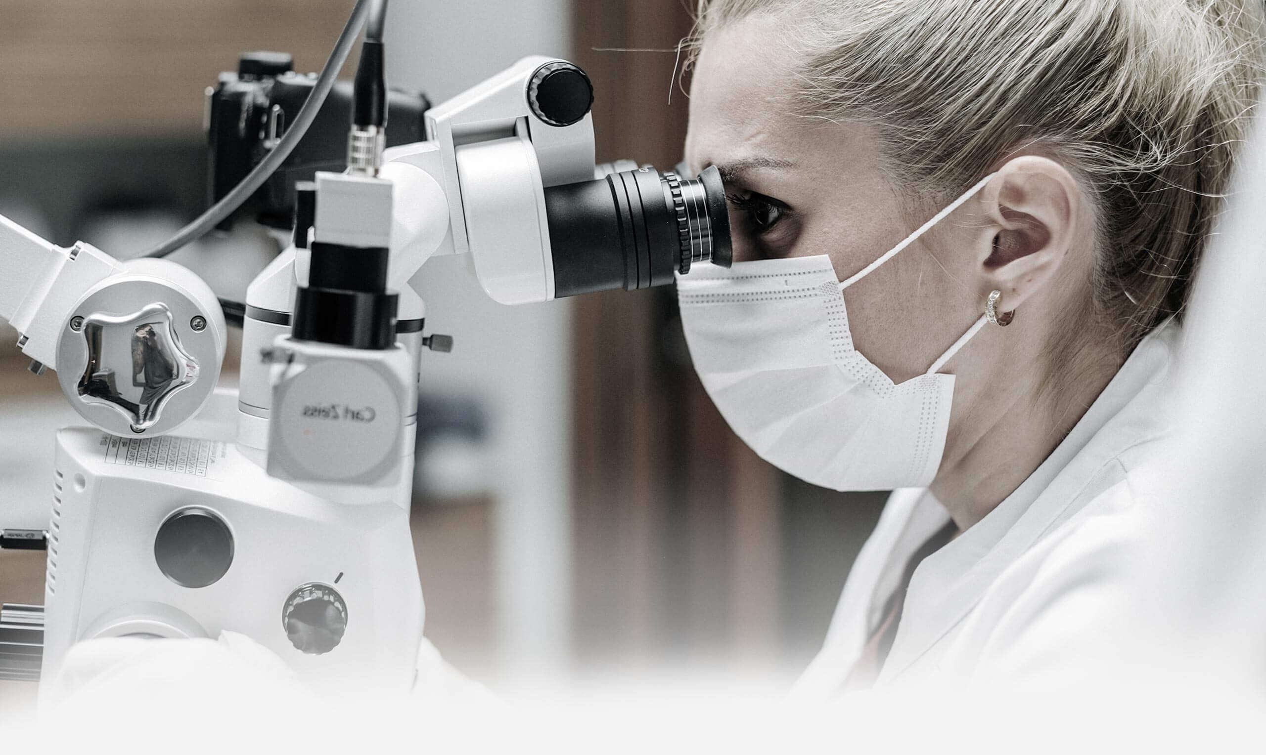
Revision of an already-present root canal treatment or root canal filling that turned out to be incomplete or insufficient or later attempt to make obliterated root canals continuous after all and to close them. Preparation, cleaning and completely reclosing a canal system. Such endodontic measures are usually performed with a dental microscope. The dental microscope or a surgery microscope permit best instrumentation of the revision or obliterated canals under view. In addition to flexible miniature drills, flexible titanium files and special endodonty ultrasound files are used for revisions. We at BERLIN-KLINIK Zahnklinik work with dental microscopes of different manufacturers when required for revisions, or with a modified surgery microscope from vascular surgery that offers about 30-fold magnification.
Inner exploration of the tooth in root canal treatment is mainly about cleaning and filling the more or less branching canal system of the tooth as gently, comprehensively and efficiently as possible. Complete internal cleaning will not be possible even with the best instruments and lenses, since the root top area often contains branches that cannot be reached and that cannot be completed, presented or prepared and filled in a controlled manner. The first technical obstacle for successful root treatment is general accessibility and visibility of this fine canal system that is partially grown shut, narrowed or wound. BERLIN-KLINIK ENDODONTIE does not want to grind off your tooth too much or drill it open too far. Surgery microscope and dental microscope are helpful for best overview. Strong grinding of the tooth and large opening of the canals are intended to force success of the measure. However, this assumption ignores an important detail: The basic question of the substantial condition of the tooth after treatment: will it be a stable, high-quality pillar, or a fragile root shard stuffed full of large amounts of root filling material?
BERLIN-KLINIK ENDODONTIE works with technical aids like ultrasound, ozone and laser, but also with high tech lenses like surgery microscopes and specialised dental microscopes. In the end, we also work with digital 2D and 3D imaging diagnosis procedures down to high-resolution, narrow-focus DVT. Digital volume tomography is already used at BERLIN-KLINIK ENDODONTIE as an aid for internal exploration of the tooth canal system. In spite of dental microscopes and laser root canal treatment, healing and prognosis are still limited by anatomy and findings that justify critical consideration of endodontic therapy in some cases. Endodonty does NOT mean that every tooth destroyed or rotted to the edge of the bone with bad roots is only kept in the jaw because we bought a dental microscope or want to prove to you that we at BERLIN-KLINIK can use the DVT to look even into difficult to view canals with our microscope! Generally, endodonty is only sensible when a tooth valuable in the long term, with maximum inner and outer substance preservation and solid healthy or partially healthy crown is the result! Over-instrumentation and over-therapy of less valuable root stumps are considered highly critical to negative at the BERLIN-KLINIK Zahnklinik. Dead teeth are, after all, dead, previously alive tissues. Even best root treatment will leave dead tissue that supports a much less beneficial metabolism than live or sterile synthetic tissue or material.
Often, particularly critical patients refuse root-treated, i.e. dead teeth, in the mouth entirely and demand sterile, stable and secure titanium implants, ideally right after extraction as immediate implant. Endodontic tooth preservation to the last stub can cause root residue that is very difficult to remove from the bone at a later stage, during surgical intervention or restoration. Teeth that have been dead for a time lose their natural retention mechanism and can grow into the jaw or wholly or partially adhere to the tooth socket wall. This ancylosis may lead to having to break or mill the extensively endodontically treated tooth from the former tooth socket alveola, causing no little difficulty. This is particularly unpleasant if it leads to the loss of important bone structure around the tooth, which may cause aesthetic degradation.
